

Pulmonary Arterial Blood Supply
Lungs Have a Dual Arterial Blood Supply (Insights Imaging, 2020) [MEDLINE]
Pulmonary Arteries
- Function
- Pulmonary Arteries Predominantly Supply the Lung Parenchyma
- Size/Pressure
- Pulmonary Arteries are Larger Caliber than the Bronchial Arteries
- Pulmonary Arteries are a Low-Pressure System
- Consequently, Most Episodes of Hemoptysis Originate from the Pulmonary Arterial Circulation and are Not Life-Threatening
- Anatomy
- Pulmonary Arteries Originate from the Right Ventricle
- Source of Hemoptysis (see Hemoptysis)
- The Bronchial Arterial System is the Predominant Source of Hemoptysis in 90% of Cases of Massive Hemoptysis (Crit Care Med, 2000) [MEDLINE]
- Other Sources Include
- Pulmonary Arteries (5% of Cases)
- Non-Bronchial Systemic Arteries (5% of Cases)
- Other Sources Include
- The Bronchial Arterial System is the Predominant Source of Hemoptysis in 90% of Cases of Massive Hemoptysis (Crit Care Med, 2000) [MEDLINE]

Bronchial Arteries
- Function
- Bronchial Arteries Predominantly Supply the Tracheobronchial Tree, Large Blood Vessels, Lymph Nodes, Esophagus, and Pleura (Radiographics, 2006) [MEDLINE]
- The Bronchial Arterial Circulation Accounts for Only 2% of the Total Vascular Supply to the Lung
- Bronchial Arteries are Very Plastic, Potentially Increasing Their Blood Flow from 1% to 30% of the Cardiac Output in Response to a Pulmonary Insult
- Consequently, Bronchial Artery Hypertrophy and Dilatation of Thin-Walled Distal Bronchial-to-Pulmonary Artery Anastomoses May Occur
- Bronchial Arteries Predominantly Supply the Tracheobronchial Tree, Large Blood Vessels, Lymph Nodes, Esophagus, and Pleura (Radiographics, 2006) [MEDLINE]
- Size/Pressure
- Bronchial Arteries Have a Smaller Caliber than the Pulmonary Arteries
- On Contrast-Enhanced CT, They are Very Thin and Difficult to Detect
- Normal Caliber of the Bronchial Arteries is <1.5 mm Near the Origin and <0.5 mm Distally, as They Branch in the Pulmonary Hila
- When Bronchial Artery Hypertrophy Occurs, Their Diameter Can Exceed 2 mm, and They Tend to Have a More Tortuous Course
- Bronchial Arteries are a High-Pressure System (Under Systemic Arterial Pressure)
- Bronchial Arteries Have a Smaller Caliber than the Pulmonary Arteries
- Anatomy
- Orthotopic Origin
- The Bronchial Arteries Most Commonly Originate from the Descending Thoracic Aorta, Usually at the T5-T6 Vertebral Plane, 1–2 cm Above or Below the Level of the Carina
- The Orthotopic Right Bronchial Artery Originates Preferentially from the Posteromedial Aortic Wall (Directly or or More Commonly from a Short Intercostal-Bronchial Trunk), Running Retrotracheally Towards the Hilum
- The Orthotopic Left Bronchial Artery Originates Preferentially from the Anterior or Lateral Aortic Wall, Running Retrotracheally Towards the Hilum
- Most Patients Have 1-2 Bronchial Arteries on Each Side and a Total of 3-4 Bronchial Arteries
- Ectopic Origin
- In Up to 36% of Cases, the Bronchial Arteries Ectopically Originate from Aortic Arch, Subclavian Artery, Thyrocervical Trunk, Internal Mammary Artery, or Coronary Arteries (J Thorac Imaging, 2003) [MEDLINE] (Eur Radiol, 2007) [MEDLINE]
- Ectopic Bronchial Arteries are Recognized Due to Their Adjacent Course with the Associated Bronchi
- In Up to 36% of Cases, the Bronchial Arteries Ectopically Originate from Aortic Arch, Subclavian Artery, Thyrocervical Trunk, Internal Mammary Artery, or Coronary Arteries (J Thorac Imaging, 2003) [MEDLINE] (Eur Radiol, 2007) [MEDLINE]
- Bronchial Arteries Have Distal Microvascular Anastomoses Connecting to the Pulmonary Arterial System (Chest, 1972) [MEDLINE]
- Orthotopic Origin
- Source of Hemoptysis
- Despite the Bronchial Arteries Accounting for Only 2% of the Total Vascular Supply to the Lung, the Bronchial Arteries are the Predominant Source of Hemoptysis in 90% of Cases of Massive Hemoptysis (Crit Care Med, 2000) [MEDLINE]
- Other Sources Include
- Pulmonary Arteries (5% of Cases)
- Non-Bronchial Systemic Arteries (5% of Cases)
- Other Sources Include
- Despite the Bronchial Arteries Accounting for Only 2% of the Total Vascular Supply to the Lung, the Bronchial Arteries are the Predominant Source of Hemoptysis in 90% of Cases of Massive Hemoptysis (Crit Care Med, 2000) [MEDLINE]
Other Arteries
- The Non-Bronchial Arterial Circulation (Aorta, Intercostal Arteries, Coronary Arteries, Thoracic Arteries, Axillary Arteries, Subclavian Arteries, Upper and Lower Inferior Phrenic Arteries) Can Supply the Lungs in <5% of Cases (Chest, 2008) [MEDLINE] (Respiration, 2010) [MEDLINE]
- Many Chronic Inflammatory Lung Lesions are Jointly Supplied by Hypertrophied Bronchial Arteries, as Well as by Collateral Systemic Arteries

Pulmonary Venous Drainage
- Bronchial Veins: uncommonly affected by pulmonary vasculitis
- Pulmonary Veins: typically located in the intralobular septa
Respiratory Muscles
- Diaphragm: innervated by C3–5
- Inspiratory Accessory: external intercostals, scalene, and sternocleidomastoid muscles
- Expiratory Accessory: internal intercostals and abdominal muscles
Airways
Definition of Airways
- Conducting Airway: extends from the trachea to the terminal bronchioles
- Functions to filter, humidify and heat air
- Respiratory Airway: includes the respiratory bronchioles, alveolar ducts, and sacs
- Site of gas exchange
- Acinus: portion of lung supplied by a primary respiratory bronchiole
Cartilage Composition of Airways
- Trachea: C-shaped cartilage with dorsal smooth muscle
- Main Bronchi: semicircular cartilage
- Bronchi: irregularly shaped cartilage plates
- Bronchioles: no cartilage support, surrounded by muscular layer
Components of Alveolar-Capillary Surface
- Surfactant
- Alveolar Epithelium: type 1 and type 2 alveolar cells (the latter of which produces surfactant)
- Interstitium
- Endothelium
Influence of Particle Size on Airway Deposition
- Particles 2–5 μm in Size: reach small airways
- Particles 5–10 μm in Size: impact on the carina or main bronchi
- Particles >10 μm: stopped in the upper airways
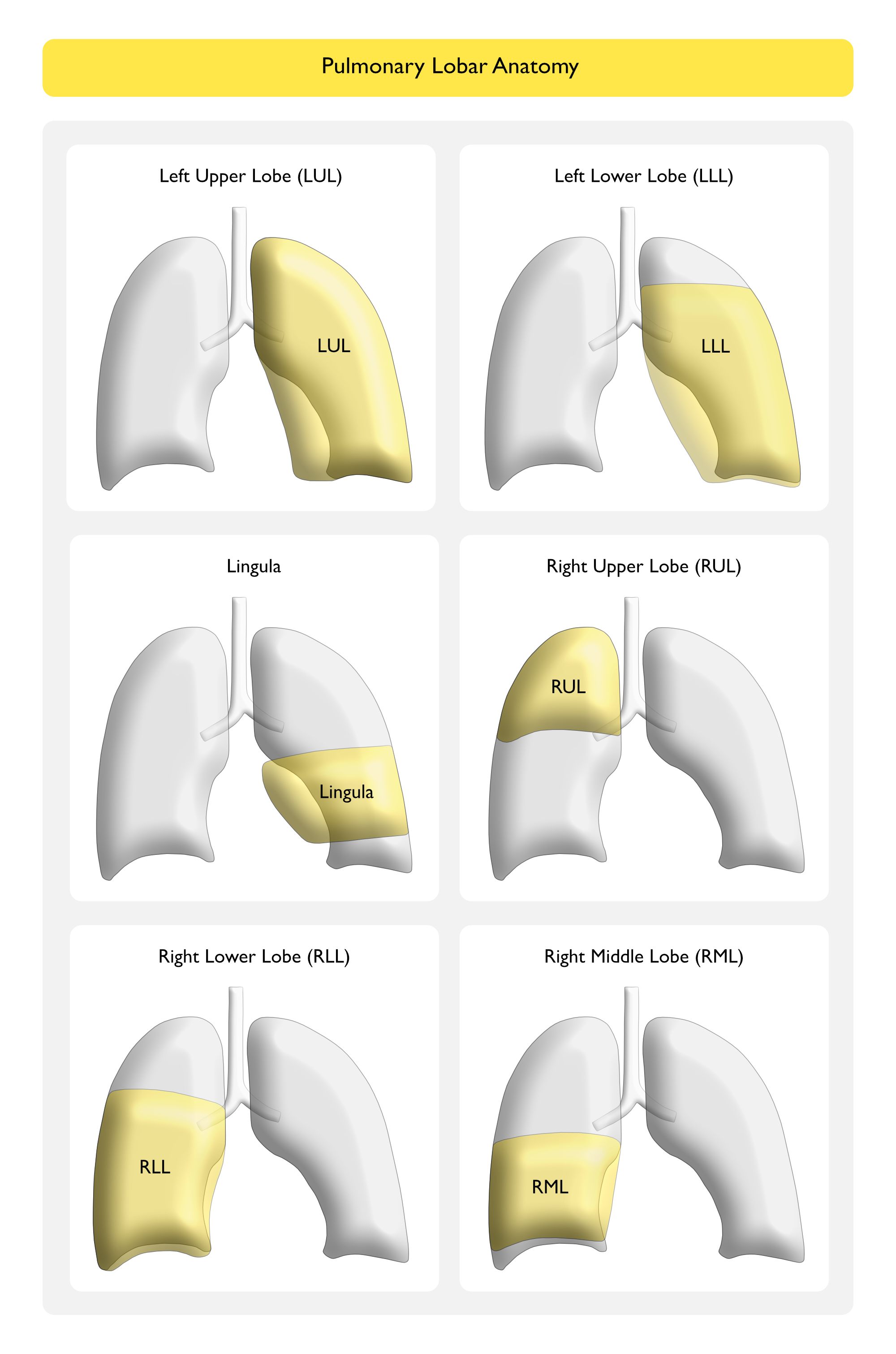
Lobar Anatomy (Frontal View of Chest X-Ray)
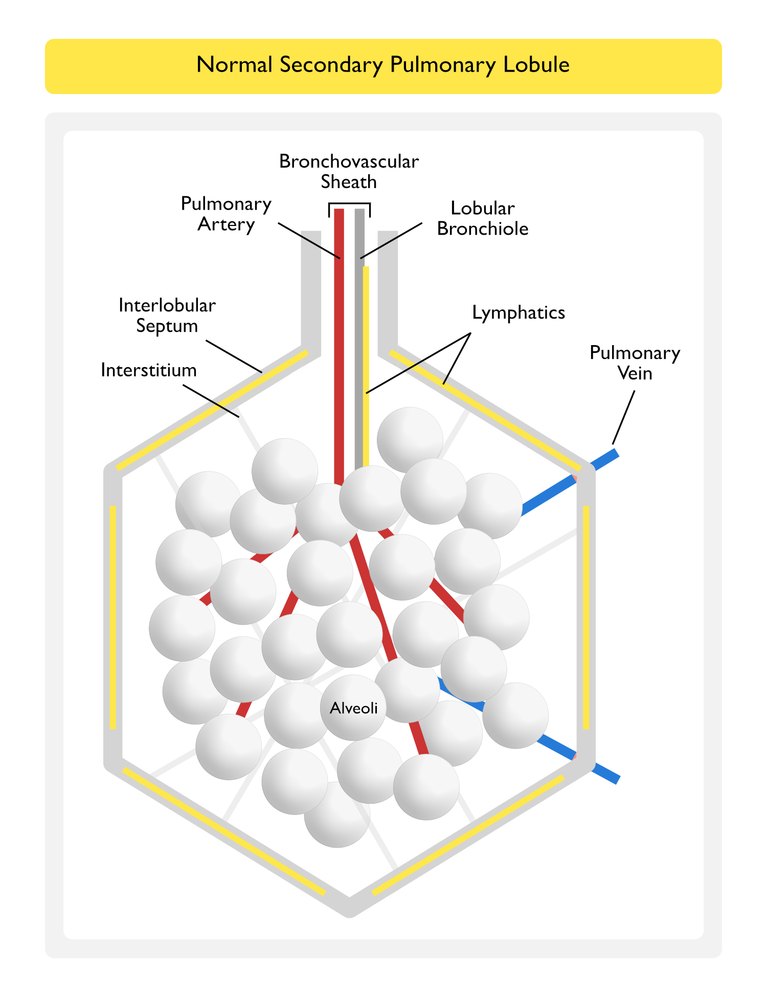
Secondary Pulmonary Lobule
The Secondary Pulmonary Lobule is a Functional Unit of the Lung Surrounded by an Interlobular Septum (W.S. Miller [The lung. 2nd ed. Springfield, IL: Charles C Thomas, 1947]) (NEJM, 2020) [MEDLINE]
- The Lobular Bronchiole and a Pulmonary Artery Branch Supply Multiple Acini within a Pulmonary Lobule
- Lymphatics and Pulmonary Veins are Located within the Interlobular Septum
- Lymphatics Also Surround the Bronchovascular Sheath
Ground-Glass Infiltrates Occur on the Chest CT Scan When Air within the Acini in the Pulmonary Lobule is Displaced by Any of the Following Four Mechanisms
- Partial Alveolar Collapse (Atelectasis) (see Atelectasis)
- Due to Obstruction, Compression, etc
- Partial Alveolar Filling (Consolidation)
- Due to Blood
- Diffuse Alveolar Hemorrhage (DAH) (see Diffuse Alveolar Hemorrhage)
- Due to Pus
- Community-Acquired Pneumonia (CAP) (see Community-Acquired Pneumonia)
- Due to Water
- Pulmonary Edema (see Pulmonary Edema)
- Near Drowning (see Near Drowning)
- Due to Cells
- Lepidic-Predominant Lung Adenocarcinoma (Previously Known as Bronchioloalveolar Cell Carcinoma) (see Lung Cancer)
- Due to Blood
- Interstitial Thickening
- Due to Interstitial Lung Disease (ILD) (see Interstitial Lung Disease)
- Thickened Interstitium
- Due to Interstitial Pulmonary Edema (see Pulmonary Edema)
- Lymphatic Engorgement by Fluid
- Due to Lymphangitic Spread of Cancer (see Lung Cancer)
- Lymphatic Engorgement by Tumor Cells
- Due to Interstitial Lung Disease (ILD) (see Interstitial Lung Disease)
- Increased Pulmonary Capillary Blood Flow

References
- Miller WS. The lung. 2nd ed. Springfield, IL: Charles C Thomas, 1947
- Evaluation of the bronchial arteries: normal findings, hypertrophy and embolization in patients with hemoptysis. Insights Imaging. 2020 May 19;11(1):70. doi: 10.1186/s13244-020-00877-4 [MEDLINE]
- Case 25-2020: A 47-Year-Old Woman with a Lung Mass. N Engl J Med. 2020 Aug 13;383(7):665-674. doi: 10.1056/NEJMcpc2004977 [MEDLINE]